This is an example of typical absence seizures (petit-mal).It is a type of generalized epilepsy. In typical absence seizure there is sudden brief loss of consciousness with cessation of speech and motor activity. The face looks blank with staring look. It is not preceded by aura and not followed by sequele. It is usually precipitated by hyperventilation or photic stimulation. With puberty it improves.
To know more visit this link:
http://www.emedicine.com/NEURO/topic3.htm
Video example of absence seizures
online virtual skeleton
If you do not have a real skeleton , this virtual one is useful for you.
This site is amazing, you can preview any bone from all views, and when pointing to a part of it , it gives you the name of this part.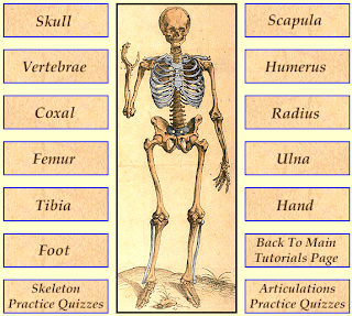

Click here to go
History and Examination at a Glance
This popular title in the at a Glance series focuses on the core elements of history taking and examination skills. History and Examination at a Glance is an ideal companion to clinical attachments and assists in building skills which are essential for passing Final Examinations and which act as a cornerstone for all medical practice. This edition features:Highly visual presentation and full colour illustrations.The latest evidence-based information to provide scientific rationale for methods usedCoverage of new topics, including the history and examination of patients with osteoporosis and HIV/AIDsNew self-assessment framework for measuring performance in history and examination skillsHistory and Examination at a Glance will appeal to all medical students as a companion throughout their clinical years, and as an essential revision aid for Final Examinations.
selected Pages:
To buy this book:
22 "must see" diagnostic radiologic images for medical students
Wonderful video from the wonderful site "Learning radiology".
You should watch it.
click on the picture to watch
Good site: Learning radiology
LearningRadiology.com is an award-winning educational site aimed primarily at medical students and radiology residents-in-training, containing lectures, handouts, images, Cases of the Week, archives of case quizzes, flashcards of differential diagnoses and “most commons” lists, primarily in the areas of chest, GI, cardiac, and bone radiology.
click the picture to go
Powassan encephalitis case
From *the Department of Medicine, McMaster University, and  the Hamilton General Hospital, Hamilton, Ont.
A previously healthy, 64-year-old man presented on Sept. 7, 1997, with a history of headache for the past 3 days and fever (38.9°C) for the past 2 days; he had also been experiencing drowsiness and slurred speech for the past day. While camping in Algonquin Park 2 weeks before presentation the patient was bitten on the buttock by an unidentified insect. The family was concerned for a number of reasons - deer mice had been reported in the park; the family cat had accompanied the patient to the family cottage in Magnetowan, Ont., and the patient had worked under the cottage prior to camping.
On presentation the patient was oriented but drowsy and slow to respond, his temperature was 38.6°C, blood pressure 105/70 mm Hg, pulse rate 90 beats/min and respiratory rate 20 breaths/min. On neurological examination significant expressive and nominal dysphasia were noted. Mild right facial weakness was present, but there was no neck stiffness. Fine rapid movements of both hands were clumsy. Muscle tone, power, sensation and muscle stretch reflexes were normal. Initial investigations showed his leucocyte count was 12.6 x 10⁹/L, predominantly neutrophils. Serum electrolytes, blood urea nitrogen, creatinine and glucose levels were normal, and the international normalized ratio (INR) and partial thromboplastin time (PTT) were normal. A chest x-ray showed early right lower-lobe consolidation. A CT scan (without contrast) of the head was normal. A lumber puncture revealed clear fluid, normal pressure, leukocytes < face="ariel,helvetica">x 106, segmented neutrophils 65%, lymphocytes 32%, monocytes 3%, glucose 3.1 (normal range 2.8-4.4) mmol/L and total protein 1.75 (normally <> admitted with a provisional diagnosis of viral encephalitis with possible brain abscess and was started on intravenous ceftriaxone (2 g iv every 12 hours) and acyclovir (500 mg iv every 8 hours). The next day a repeat CT scan (with contrast) of the head was normal, and an echocardiogram was negative. Two days after presentation (Sept. 9) the patient began to experience right side weakness and a decreased level of consciousness, with no response to verbal or tactile stimuli. His right pupil was slightly more dilated than his left and muscle tone was flaccid, with muscle stretch reflexes 1+. The patient could not protect his airway and was intubated and transferred to the intensive care unit. Laboratory results of blood taken for viral serology (for eastern equine, western equine, St. Louis and Powassan virus antibodies and Hantavirus, rabies and Lyme disease) were negative. The following day (Sept. 10) an MRI brain scan was normal, and a lumbar puncture showed clear fluid, normal pressure, leukocytes 113 x 106, lymphocytes 60%, glucose 3.5 mmol/L and total protein 0.83 g/L. An EEG showed diffuse slowing and disorganization, and a diagnosis of viral encephalitis was established. Eight days after presentation (Sept. 15) the patient began to have episodes of awakening and was more responsive to pain and verbal requests. On September 18, 11 days after presentation, blood viral serology was positive for Powassan antibody (1/160). By September 23, the patient was more responsive; he could open his eyes and follow objects, but he was still weak and areflexic. Another CT scan (with contrast) performed on October 1 was normal. One month after presentation (Oct. 7) facial expressions were evident, and the patient was able to elevate his shoulders and bend his knees.
On Oct. 16, 1997, while sitting for physiotherapy, the patient suffered a cardiac arrest and could not be resuscitated. The cause of death determined at autopsy was a massive pulmonary embolism. The patient had received subcutaneous heparin (500 U twice daily) for deep vein thrombosis prophylaxis. Neuropathologic examination showed mild diffuse swelling of the cerebral hemispheres with diffuse meningeal congestion. Histological examination revealed an intense chronic inflammatory infiltrate in the meninges and Virchow-Robin spaces, with focal areas of infiltration into brain parenchyma in the most severe areas associated with tissue necrosis. Areas most involved were the mediotemporal lobes, ventral midbrain and basal ganglia. There was no vasculitis or true infarction. Cerebral white matter was clearly less affected and largely unremarkable, aside from secondary changes and edema. Throughout the grey matter there was diffuse reactive astrocytic gliosis, as well as microglial activation. Careful examination showed the occasional neuron with an intranuclear eosinophilic inclusion, likely representing a viral inclusion. No viral particles or inclusion structures were seen upon electron microscopic examination of brain tissue taken at the time of autopsy, although the examination was limited by sampling and postmortem artifact.Read more ......
WHAT IS THE DIAGNOSIS? : a good site
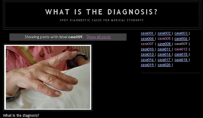
Very good useful site for medical students.It presents cases in the form of spot diagnosis.Really good site , you should visit.Click here to go
Rene and Weber tests
Movies drawn from the Neurologic Exam and PediNeurologic Exam websites are used by permission of Paul D. Larsen, M.D., University of Nebraska Medical Center and Suzanne S. Stensaas, Ph.D., University of Utah School of Medicine. Additional materials for Neurologic Exam are drawn from resources provided by Alejandro Stern, Stern Foundation, Buenos Aires, Argentina; Kathleen Digre, M.D., University of Utah; and Daniel Jacobson, M.D., Marshfield Clinic, Wisconsin. Subsequent re-use of any materials outside of this program, presentation, or website requires permission from the original producers.
physical examination: Loyola University Chicago
Full female examination from head to toe (wmv)
http://rapidshare.com/files/97287962/full-female-exam-1of4.rar.html
http://rapidshare.com/files/97248488/full-female-exam-2of4.rar.html
http://rapidshare.com/files/96388685/full-female-exam-3of4-wmv.rar.html
http://rapidshare.com/files/96373180/full-female-exam-4of4-wmv.rar.html
Password: 4medstud.blogspot.com
Subscribe to:
Posts (Atom)